On October 19, 2021, Assistant Professor He Shuijin’s group at SLST published a research article entitled “PUPIL enables mapping and stamping of transient electrical connectivity in developing nervous systems” in Cell Reports. In this study, they modified PUP-IT (developed by SLST Assistant Professor Zhuang Min, (read more at https://www.shanghaitech.edu.cn/eng/2018/0815/c1418a31492/page.htm) to enable the pupylation-based interaction labeling (PUPIL) of electrical synapses in vivo and found that PUPIL can noninvasively and efficiently map and stamp transient CX26 that contains electrical synapses in the mouse neocortex.
Electrical synapses are distributed in multiple tissues, including the nervous system, cardiovascular system, and reproductive system, and play important roles in development, synaptogenesis and neuronal synchronization. Conventional methods for assessing electrical connectivity and function include dye injection, paired electrophysiological recordings, and electromicroscopy imaging, all of which are inefficient, invasive, and laborious for mapping overall electrical connectivity. Researchers over the past decades have focused on mapping neuronal chemical connectivity, prompting development of a number of relevant genetic tools such as green fluorescent protein (GFP) reconstitution across synaptic partners (GRASP) and trans-synaptic neuronal tracing. However, genetic methods for mapping in vivoelectrical connectivity remain elusive.
In this study, the researchers applied PUPIL to map and stamp electrical synapses to investigate the role of electrical synapses in cortical neuron development. They validated that PUPIL detected actual electrical synapses, but not artifact substrate aggregate, with immunostaining, electron microscopy and paired whole-cell recordings. In combination with the GFP reconstitution or GCaMP approaches, PUPIL can be used to image electrical synapses in living tissues and reveal their precise subcellular localizations in the mouse neocortex. Moreover, they applied PUPIL to elucidate physiological functions of transient CX26. Additionally, they applied PUPIL to label CX36, containing gap junctions that primarily form among interneurons in the brain, and label the inhibitory synapses, suggesting that PUPIL has great potential to track the dynamic behavior of chemical synaptic dynamics.
Third year Ph.D. candidate Xie Shu is the first author, and Prof. He is the corresponding author. The research is supported by the Molecular Imaging Core Facility of SLST, Bio-Electron Microscopy Facility of ShanghaiTech and Electron Microscopy Core Facility of Center for Excellence in Brain Science and Intelligence Technology, Chinese Academy of Sciences.
Link to the article: https://www.sciencedirect.com/science/article/pii/S2211124721013206
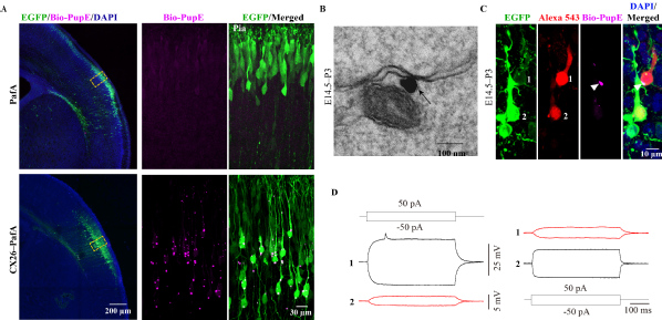
Biotin signals are verified by antibody immunostaining, immunoelectron microscopy and electrophysiological recording