SIAIS Principal Investigator Dr. Rao Zihe and his team recently unveiled the 3-dimensional structure of the African swine fever (ASF) virus, a giant and complex virus which causes a contagious and lethal swine disease all over the world. The study, entitled ‘Architecture of African swine fever virus and implications for viral assembly,’ was published in Science on Oct 17th.
Since first described in Kenya in 1921, ASF virus has spread through many countries including China. Without vaccine or effective treatment available, the ASF virus has caused huge economic losses worldwide. The 3-dimensional structure of the ASF virus sheds light on the mechanism of the disease and subsequently provides clues for vaccine development.
In this work, the research team constructed the ASF virus capsid structure up to 4.1-angstroms, using the cryo-electron microscopy (EM) method. The structure includes five layers: the nucleoid, the core shell, an inner lipid envelope, an icosahedral protein capsid, and an external envelop. With a diameter of 260-300 nm, 10 times longer than that of the hepatitis A virus, the ASF virus structure built from 17280 proteins is the largest among virus structures solved by Chinese scientists.
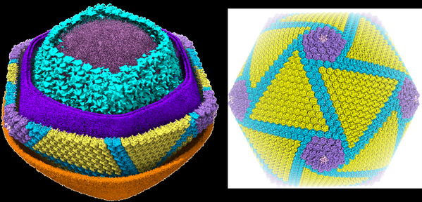
The architecture of African swine fever virus
Due to its large size, the structure is significantly more complicated with the ASF virus than with other viruses, requiring a large amount of high-resolution EM data. The access for cryo-EM instrument became a bottleneck at first. Considering that the ASF virus study was of urgent need in our country, ShanghaiTech University provided full support to the study, granting Dr. Rao’s team special access to the Bio-Electron Microscopy Facility of ShanghaiTech University from February to May 2019 during the facility’s trial operation. The team spent four months at the facility collecting cryo-EM data, which is almost impossible to do at any other EM facilities. With this support, Dr. Rao’s team effectively collected over 100 TB of cryo-EM data with over 70,000 high-quality images, from which the 3-dimentional structure was successfully constructed. The support from the Bio-Electron Microscopy Facility of ShanghaiTech University was vital and crucial to the team’s successful accomplishment of the ASF virus structure.
The Bio-Electron Microscopy Facility was implemented in August 2018 and opened for trial operation in December 2018, to meet the needs of researchers for electron microscopy. It has now become one of the most advanced electron microscopy facilities in China. The facility currently houses five electron microscopes, including two Talos 120C, one medium Arctica (200kv) and two Titan Krios (300kv). It also provides sample preparation instruments, including CLEM CorrSight, High Pressure Freezing, cryo-ultramicrotomy, and GPU-based high-performance computing cluster resources, to provide comprehensive support for cryo-EM data collection, storage and analysis.
With state-of-the-art cryo-EM instrumentations, the Bio-Electron Microscopy Facility of ShanghaiTech University provides researchers access to cutting-edge technology for high resolution imaging of subcellular and biological macromolecules. As a key component of the public service platform, the Bio-Electron Microscopy Facility of ShanghaiTech University will serve users from academia, healthcare as well as industry, accelerating the development of biomedical research, drug discovery and technology transfer.
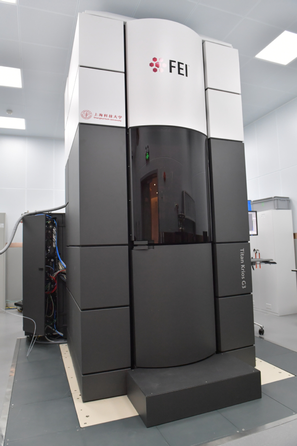
The Bio-Electron Microscopy Facility of ShanghaiTech University
(In the picture: Titan Krios (300kv) electron microscopy)
The study was delivered jointly by Dr. Rao’s team and collaborators from the Institute of Biophysics of the Chinese Academy of Sciences (CAS), the Harbin Veterinary Research Institute of the Chinese Academy of Agricultural Sciences (CAAS), ShanghaiTech University and other institutes. The cryo-electron microscopy (EM) data collection in this study was conducted at the Bio-Electron Microscopy Facility of ShanghaiTech University.
Read more at: https://science.sciencemag.org/content/early/2019/10/16/science.aaz1439.full