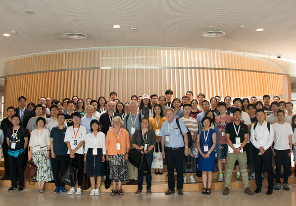
From July 30th to 31st, the Forum on Imaging Across Scales and Cell Mapping and the Cryo-EM Facility Inauguration Ceremony of ShanghaiTech University was successfully held at the iHuman Institute of ShanghaiTech University. Chaired by Professor Raymond Stevens, the forum hosted more than 100 participants including scholars, researchers and students from the National Center for Protein Science Shanghai (NCPSS), University of Science and Technology Beijing, Nantong University, Tsinghua University, Shanghai Jiaotong University, Tongji University, Westlake University, Zhejiang University, Shanghai Institute of Materia Medica of Chinese Academy of Sciences (CAS), Shanghai Institute of Biochemistry and Cell Biology of CAS, Shanghai Institute of Organic Chemistry of CAS, Nankai University, and other academia in China and abroad.
Over the past two decades, we have experienced breakthrough developments in multi-scale biological imaging techniques including optics, acoustics, mass spectrometry, electron microscopy, X-ray tomography, nuclear magnetic resonance, and many others. These advanced techniques allow us to observe and characterize the static structures, dynamic processes, molecular networks, cellular neighborhoods, entire cells and organs. This forum aims to identify the perspectives, challenges, and opportunities of imaging across scales, and to review and push forward the cell mapping at iHuman Institute regarding pancreatic beta-cells, neurons, and organs. This forum invited distinguished international guest speakers in the research field of bio-imaging, guest speakers from Tsinghua University, Center of Cryo-Electron Microscopy of Zhejiang University, iHuman Institute and School of Information Science and Technology (SIST), experts in bio-mass spectrometry of Shanghai Institute of Materia Medica of CAS and image processing scientists from Thermo Fisher Scientific.
The forum consisted of both oral talks and panel discussions. Professor Raymond Stevens (iHuman Institute, ShanghaiTech University/University of Southern California) gave the opening remarks, welcomed the guests and scholars, and introduced the progress and collaborations of iHuman Institute with other research institutions regarding imaging across scales. He also gave a brief introduction to the history, the research field, and interdisciplinary features of the forum. Professor Wolfgang Baumeister (ShanghaiTech University/Max Planck Institute of Biochemistry, Germany) presented the developments and achievements of cryo-electron tomography, and highlighted its unique potential for imaging macromolecular polymeric proteins in functional cellular environments. Professor Kate White (University of Southern California) introduced the Pancreatic β-Cell Consortium, the current application of soft X-ray tomography in investigating the mesoscale atlas of β-Cell and its significance in whole-cell modeling. Professor Carl Kesselman (University of Southern California) introduced the data organization and sharing framework in Pancreatic β-Cell Consortium and shared how we can better use data to drive collaboration and accelerate research. Professor Helen Berman (Riggers University/University of Southern California) showed the management of integrated protein structure models in protein data bank. Professor Andrej Sali (ShanghaiTech University /University of California, San Francisco) introduced the integrative modeling of nuclear pore complexes and emphasized the significance and development of the integrative modeling framework.
After the plenary talks, all attendees visited SIST’s Multi-disciplinary Artificial Reality Studio (MARS). Through the form of a hologram, SIST Vice Dean Professor Yu Jingyi introduced the application of machine learning methods in full-body 4D holographic imaging and other cutting-edge researches. In addition, experts from the CAS and Thermo Fisher Scientific introduced how to use the hydroquinone exchange mass spectrometry combined with cryo-electron microscopy to analyze the protein structure, how the MS tool box can help to clarify the foggy spot on atomic resolution cryo-EM protein structure, as well as the new technologies in cryo-electron microscopy.