A team of scientists led by Professor Huaidong Jiang and Professor Zhi Liu of the School of Physical Science and Technology at ShanghaiTech University, along with Professor Chunying Chen of the National Center for Nanoscience and Technology of China, Professor Renzhong Tai of the Shanghai Institute of Applied Physics have made important progress in the field of bioimaging based on synchrotron radiation facility. The researchers performed multi-model imaging techniques such as scanning transmission X-ray microscopy, X-ray fluorescence microscopy, and combined with new nano-CT algorithms to achieve high-resolution imaging of mammalian macrophages, and quantitatively observed three-dimensional distribution of potential nanomedicine [Gd@C82(OH)22]n within the cells. Their work provides important information for understanding the high-efficiency and low-toxicity anti-tumor effect of nanomedicine. The paper was published in the March issue of IUCrJ as a cover article entitled “Three-dimensional ultrastructural imaging reveals the nanoscale architecture of mammalian cells.” A scientific commentary article entitled “Multi-model imaging of the interaction of nanomaterials with cells” was published in the same issue of IUCrJ.
Understanding interactions between functional nanomaterials and intracellular biological structures at the cellular level is crucial for studying biological effects in nanomedicine and nanotoxicology. So far, optical microscopy and electron microscopy are the most commonly used techniques for cellular studies. However, optical microscopy is limited by factors such as resolution and fluorescent labeling, and it is impossible to perform 3D imaging of intracellular nonfluorescent structures at nanometer resolution. For electron microscopy, sample preparation techniques still suffer from tedious steps and only cell slices can be observed due to the poor penetration depths of electrons. Synchrotron-based X-ray microscopy can fill the gap for structural investigation of whole cells at nanoscale by taking advantage of strong penetrability and short wavelength of X-rays.
This work was mainly carried out on the beamline BL08U1A at Shanghai Synchrotron Radiation Facility (SSRF). In this study, X-ray spectroscopy, scanning transmission X-ray microscopy and an iterative tomographic algorithm called equally sloped tomography were combined to investigate individual macrophages exposed to the potential antitumor agent [Gd@C82(OH)22]n. The macrophages grown on the carbon membrane were scanned through the focal plane of X-ray beam perpendicularly. In order to achieve 3D cell structure, 46 projections were measured at equally sloped angles within a range of ±79.4° for each data set. At the same time, according to the abrupt change in the X-ray absorption edge of a specific element (Gd) and the energy-adjustable characteristics of the synchrotron radiation source, researchers realized chemically resolved imaging of biological cells. The nanomaterials containing Gd in cells can be directly observed and accurately localized. Thus, 3D distribution of the nanomaterials can be performed and quantitatively analyzed with a resolution of 75 nm (Figure 1). This imaging technique provides an intuitive and reliable quantitative approach to the study of nanomaterials within intact large-size cells at the nanometer scale and will greatly benefit the fields of nanomedicine and nanotoxicology.
The first author of the paper is Dr. Shengkun Yao from the School of Physical Science and Technology and Professor Huaidong Jiang is the corresponding author. This study was supported by the National Natural Science Foundation of China, the Major State Basic Research Development Program of China, ShanghaiTech University and Shanghai Synchrotron Radiation Facility.
Read more at: https://journals.iucr.org/m/issues/2018/02/00/mf5022/index.html and https://journals.iucr.org/m/issues/2018/02/00/hi5652/index.html

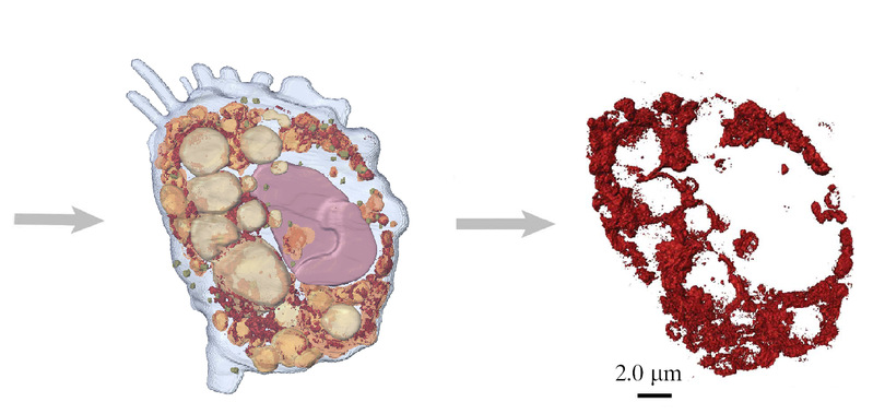
Fig. 1. 3D imaging of single cell and the distribution of intracellular nanomaterials